SMELL
Humans have a very keen sense of smell: we can detect thousands of different smells. This ability relies on the presence of special sensory receptors in the upper part of the nose. When stimulated by odour molecules, these receptors send signals along nerves to the brain for processing. Sometimes odour molecules do not reach the sensory area, but sniffing will help get them there.
SMELL RECEPTOR CELLS IN ROOF OF NOSE
Smell receptors are specialized nerve cells. Each bears many tiny cilia (hairs), which project into the space in the upper part of the nose. A nerve fibre extends from the other end of each cell. This joins other fibres to form the olfactory nerves, which carry signals to the brain.
CILIA OF SMELL RECEPTOR CELL
The cilia can detect tiny amounts of substances in the air, though molecules of those substances must first be absorbed by the mucus layer. There they interact with the cilia to trigger nerve impulses.
TASTE
We can taste substances in food and drink thanks to the 10,000 or so taste buds located on structures, called papillae, on the surface of our tongues. These receptors send signals along nerves to the brain for interpretation. Four main tastes – sweet, salty, sour, and bitter – are detected by the taste buds in four areas of the tongue. The senses of taste and smell combine to analyse flavours.
PAPILLAE ON SURFACE OF TONGUE
Papillae are tiny protrusions on the surface of the tongue. The fungiform papillae and some other types of papillae contain taste buds. The smaller, more numerous, filiform papillae do not contain taste buds but give the tongue a rough surface, which helps it move food around the mouth.
TASTE RECEPTOR CELLS IN A TASTE BUD
Hairs emerge from each receptor cell. Food and drink molecules must dissolve in saliva before they can interact with these hairs and trigger signals to the brain.
HEARING
Our ears allow us to detect sounds, which pass through the air as waves of varying pressure. On reaching the ear, the waves travel through several structures to the cochlea in the inner ear. There, receptor cells produce signals that go to the brain. The human ear can detect sounds over a very wide range of pitch and loudness, from the high-pitched squeaks of a mouse to the roar of a passenger jet.
HEARING APPARATUS OF THE EAR
The outer ear channels sound waves into the ear canal. These sound waves cause the eardrum, a thin membrane at the end of the ear canal, to vibrate. The vibrations are transmitted via three tiny bones in the middle ear to the cochlea in the inner ear.
Inside the cochlea, sound vibrations make these sensory hairs move, which triggers signals in attached receptor cells. The signals pass to the brain, which works out the pitch and loudness of the sound.
BALANCE
balance is an internal sense and relies on sensory receptors that monitor the position of the head and body. Whether we are still or moving, balance is essential for maintaining our posture and stopping us falling over. The vestibule and semicircular canals of the inner ear provide information on the position and movements of the head. Combined with signals from the eyes, this helps us balance.
BALANCE APPARATUS OF INNER EAR
Turning movements of the head are picked up by sensory hair cells embedded in structures called cupulae in the semicircular canals. Tilting movements of the head, and its position, are monitored by hair cells within structures in the vestibule.
These tiny crystals, called otoliths, are attached to sensory hair cells in the vestibule. When the head tilts, the otoliths move, causing the hair cells to bend and nerve signals to be sent to the brain.
SIGHT
Whenever we are awake, our eyes work constantly to collect information about the world. As this data is analysed by the brain, we are supplied with a detailed picture of our surroundings. We can judge distance, see in dim and bright light, and experience COLOUR VISION. For us to see, light rays reflected by objects around us must meet at the back of the eye. There they trigger electrical signals that are sent to the brain for interpretation.
The eyes sit in two bony cavities in the skull. Light rays entering the eye pass through the cornea, lens, and vitreous humour, before reaching the retina, the light-sensitive area at the back of the eye. Signals generated in the retina leave the eye along the optic nerve and go to the brain. Around each eye lie six tiny muscles, which enable the eye to turn and swivel in its socket.
Light rays from an object are refracted (bent) first by the cornea and then by the lens, which can be made to change shape according to the distance of the object from the eye. The refraction of the rays ensure that they meet on the retina. There, images are formed upside down, but the brain makes sense of this information, so we see objects the right way up.
Nerve signals leave the eyes in the optic nerves, which meet at the optic chiasma. There, fibres from the inner side of each retina cross so that each side of the brain receives information from each eye. The signals pass along the optic tracts to linked areas at the back of the brain. This part of the brain, called the visual cortex, forms a three-dimensional image of the object being viewed.
In bright light or when viewing close objects, the pupils of our eyes constrict (narrow). This is caused by tightening of circular muscles within the iris, the coloured region of tissue that surrounds the pupil. The constriction of the pupil reduces the number of light rays entering the eye
In dim light or when we are viewing distant objects, our pupils dilate (widen). This is due to tightening of a different set of iris muscles that are arranged like spokes in a wheel around the pupil. Full widening of the pupil allows the maximum number of light rays to enter the eye.
The retina houses two types of light-sensitive cells: rods and cones. The cones give us colour vision. There are three different types of cone, each sensitive to light within a different range of light wavelengths (colours). Signals are sent from the cones to the brain. From the overall pattern of signals, the brain can work out the colour of every tiny point in the scene being viewed.
HOW THE RETINA RESPONDS TO LIGHT
When light rays reach the retina, they trigger chemical changes in different light-absorbing substances in the rod and cone cells. These changes trigger electrical signals in the cells. The rods and cones link to a system of connecting nerve cells. These perform some initial processing of the signals and then transmit them along optic nerve fibres to the brain.
In each retina, the rods (seen here coloured grey) outnumber the cones (coloured orange) by about 17 to 1. The cones only work in bright light, whereas rods respond to dim light. Unlike cones, rods are all of the same type. They are responsible for the black-and- white vision we experience in semi-darkness.
TOUCH
Your sense of touch works by means of special sensory receptors scattered all over your body’s surface. These receptors allow you to feel an amazing range of sensations, from the pain of touching a searing hot iron to the tickling of a feather as it brushes against your skin. The receptors send messages along nerves to the spinal cord and brain, where the information is processed.
Touch receptors are types of specialized nerve ending. Meissner’s corpuscles detect fine touch and are found in hairless parts of the body, such as the lips, palms, and fingertips. Other types of receptor are sensitive to pressure, stretching of the skin, vibration, or hair movements.
Some areas of the skin, such as the fingertips and palms, are folded into ridges. These help improve both touch sensitivity (as they hold more receptors) and grip. The pattern of ridges and grooves provides a means of identification, because everyone has their own unique ridge pattern.
Developed in the 19th century by a Frenchman, Louis Braille, the Braille system allows blind people to read. Words are represented by a series of raised dots, which the reader recognizes by running his or her fingers over the page. The ability to read Braille relies on the extreme sensitivity of the fingertips to touch.
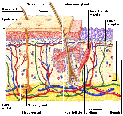
SKIN
The skin, along with hair and nails, provides the body with a protective outer covering that shields it, for example, from harmful solar rays. It also provides our first line of defence against infection, helps control water loss from the body, plays an important role in
TEMPERATURE CONTROL, and contains the receptors that provide the sense of touch.
The skin has two main layers, called the epidermis and dermis. The epidermis consists of an upper layer of dead cells and a lower living layer, which replaces cells as they are lost from the upper layer. Beneath the epidermis is the thicker dermis, which overlies an insulating layer of fatty tissue.
Hair grows from follicles, pockets of epidermal tissue that extend down into the dermis. Hair has a cycle of growth, rest, and then loss, when the new hair pushes the old hair out of the follicle. About 100 hairs are lost and replaced in a person’s scalp every day.
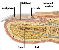
The ends of the fingers and toes are covered by nails. These plates of tough protective tissue are made mainly of keratin, a protein also found in hair and skin. Nails grow from a region of living cells called the germinal matrix, which lies underneath a fold of skin called the cuticle.
The blood vessels, hairs, and sweat glands of the skin work together to help control body temperature. If we get too hot, our sweat production increases and blood vessels widen to allow more blood to reach the skin’s surface, where it cools. If we get too cold, these processes go into reverse. In addition, tiny muscles attached to the hair follicles pull the hairs erect, trapping an insulating layer of air next to the skin.
Sweat, a salty liquid, reaches the surface of the skin through pores. The pore is surrounded by dead epidermal cells. Sweat evaporates from the surface of the skin and so helps to lower the body temperature. Sweating also rids the body of excess water and some waste products.
No comments:
Post a Comment